The exploration of neural circuits has significantly advanced our understanding of brain functionality, particularly in how neurons communicate and process information. Central to these explorations are genetically encoded voltage indicators (GEVIs), which enable scientists to visualize electrical activities within the brain. However, a critical point of discussion is the effectiveness of one-photon (1P) imaging as opposed to two-photon (2P) imaging techniques. Recent research conducted by a team from Harvard University has offered substantial insights into the advantages and drawbacks of these methodologies, significantly impacting the trajectory of future research in neuroscience.
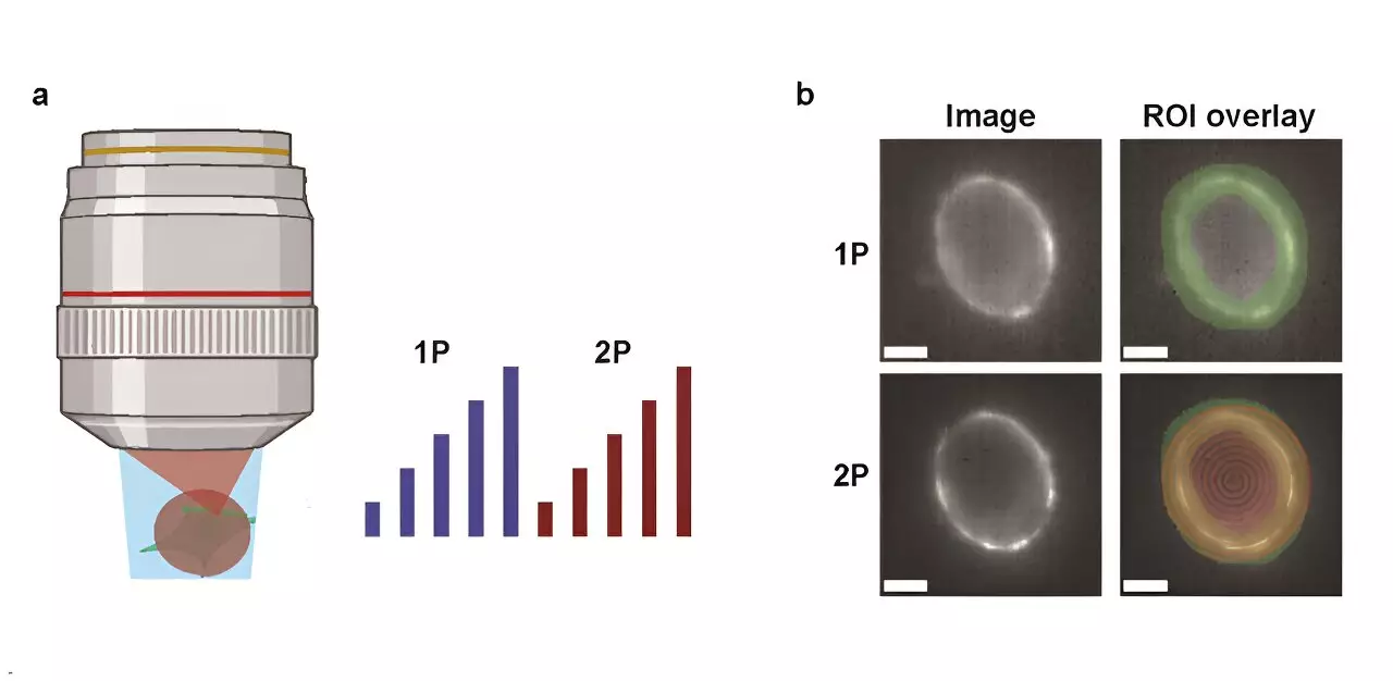
Published in the journal *Neurophotonics*, the study presents a meticulous side-by-side analysis of 1P and 2P voltage imaging, emphasizing the inherent optical and biophysical limitations each method possesses. A major aspect of the study was the evaluation of several commonly utilized GEVIs, assessing their brightness and sensitivity to voltage fluctuations under both imaging techniques. One of the critical measures taken was to gauge how fluorescence intensity diminishes as imaging depths increase, particularly within the intricacies of the mouse brain—a vital consideration for in vivo applications.
To effectively analyze their results, the researchers constructed a predictive model that estimates the number of cells identifiable based on distinct reporter properties, imaging strategies, and the essential signal-to-noise ratio (SNR). This model not only provides insight into the potential of voltage imaging but also serves as a guide for future experimentation.
Among the most revealing conclusions drawn from this research is the stark difference in illumination requirements between the two techniques. Specifically,2P voltage imaging necessitates an astonishing 10,000 times more power for each targeted cell compared to 1P methods to reach equivalent photon yield rates. This substantial energy demand introduces notable complications, particularly concerning tissue damage and the generation of shot noise—a critical element that can confound results in high-resolution imaging.
An example illustrates this challenge effectively: using the JEDI-2P indicator in the mouse cortex at specified thresholds, researchers found the capacity limited to collecting data from approximately 12 neurons beyond a depth of 300 micrometers when adhering to safe power limits and specified measurement conditions. Such constraints underscore the ongoing difficulty researchers face with 2P voltage imaging, which is hindered further by the current GEVIs’ modest voltage sensitivity.
The findings from this study paint a clear picture of the need for innovation within the realm of voltage imaging. To enhance the capability of 2P imaging and make it a viable option for recording substantial numbers of neurons deep within the brain, significant advances in current GEVIs or the invention of new imaging techniques are imperative. The juxtaposition of 1P and 2P methods illustrates not just a technical challenge but a broader narrative of how scientific tools can evolve to meet the complexities of biological systems.
As research continues to push the boundaries of neuroscience, studies such as these from Harvard will be instrumental in guiding future endeavors in imaging technologies. They highlight not only the limitations that exist but also the burgeoning potential for breakthroughs that could reshape our understanding of neural circuits. The quest for improved voltage imaging devices remains a priority for researchers aiming to decode the intricate workings of the brain, ultimately fostering greater progress in the field of neuroscience.