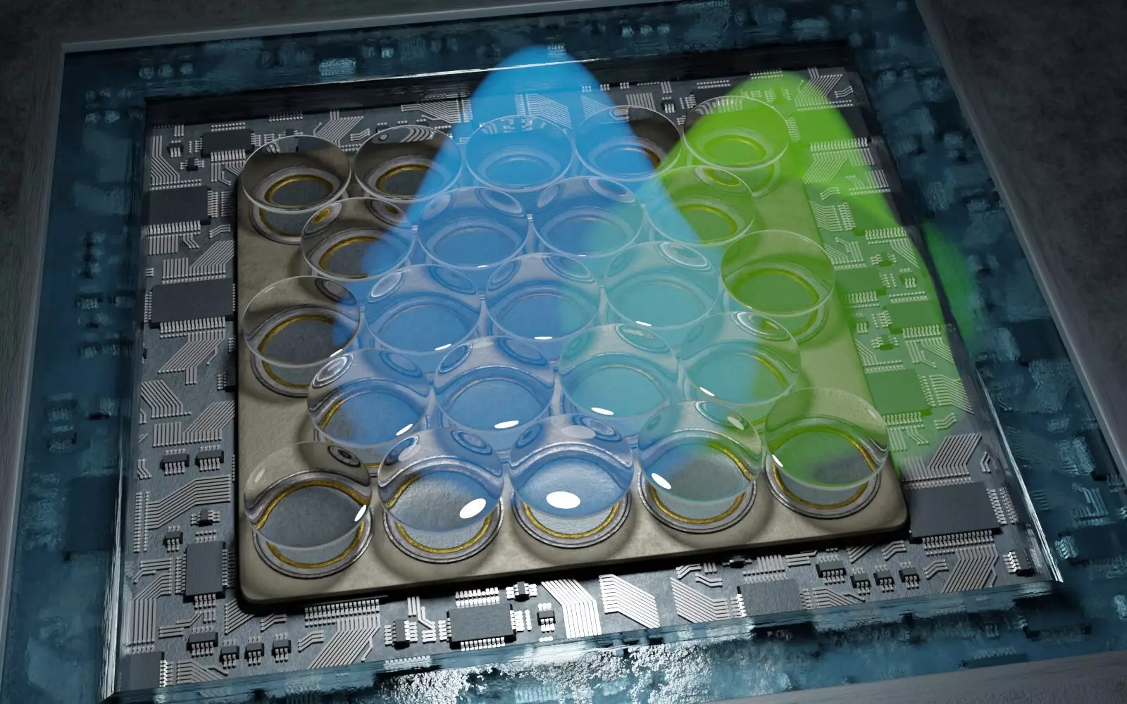
The microscopic world within our cells has long teased scientists, revealing clues only in fragments due to the limitations of conventional microscopy. Traditional techniques often struggle to unveil the intricate structures that reside at nanometer scales, leaving many cellular mysteries unsolved. However, an innovative development by researchers from the Universities of Göttingen and Oxford, in collaboration with the University Medical Center Göttingen, has ushered in a new era of cellular imaging, boasting resolutions finer than five nanometers—a feat that transforms our understanding of biology at the most fundamental level.
Historically, biological imaging has faced significant challenges, particularly when it comes to visualizing small structures within living cells. Conventional microscopes typically achieve resolutions starting at around 200 nanometers, which is inadequate for observing the minute architectural details critical to cellular function. For instance, elements of cellular scaffolding, like the fine tubes that can measure around seven nanometers in diameter, remain elusive to these traditional methods. Additionally, the synaptic clefts, the minute gaps through which nerve cells communicate, range from 10 to 50 nanometers—again, too fine to be captured adequately. This gap in resolution has limited our understanding of cellular processes such as signaling and interaction, potentially stymying progress in various fields, from neuroscience to cell biology.
The breakthrough technology developed by the Göttingen researchers is a specialized form of fluorescence microscopy, which hinges on a technique called “single-molecule localization microscopy.” This method allows for the precise tracking of individual fluorescent molecules within a sample, making it possible to model entire structures based on the exact locations of these molecules. Previously, such methodologies provided resolutions of around 10 to 20 nanometers, but the team, led by Professor Jörg Enderlein, has achieved an astonishing doubling of this resolution. Utilizing highly sensitive detectors combined with sophisticated data analysis techniques, they have refined the ability to visualize cellular components with unparalleled clarity.
To grasp the scale of this achievement, consider the analogy of measurements; one nanometer compared to one meter is akin to comparing the diameter of a hazelnut with the expansive diameter of the Earth. This comparison illustrates the immense difference that nanometer-scale imaging makes in the biological field, where understanding the roles of proteins and their organizations within cells is paramount. Professor Enderlein’s team emphasizes that these advancements are not just about the resolution itself; they represent a transformative leap in high-resolution microscopy that renders it both more accessible and cost-effective compared to existing techniques.
As scientists unlock the finer details of protein interactions and cellular architecture, potential applications abound. Researchers can delve deeper into neuroscience, studying how neurons communicate and how synaptic organization influences brain function. In cancer research, improved imaging may unveil how tumor cells migrate or how they interact with surrounding tissues. This level of detail could dramatically influence therapeutic approaches.
In addition to enhancing imaging capabilities, the team’s commitment to accessibility is noteworthy. They have developed an open-source software package for data processing, broadening the availability of this advanced microscopy to researchers across diverse disciplines. This democratization of technology promotes collaborative advancements in cell biology, biochemistry, and several other fields that rely on high-fidelity imaging.
The impact of this innovation transcends its technical prowess; it signifies a burgeoning era where even the tiniest cellular structures may no longer be shrouded in mystery. As scientific inquiry continues to push boundaries, this new technology will likely inspire further research, prompting fresh hypotheses and facilitating discoveries that may redefine our understanding of life at its most intricate scale.
The development of a microscope with nanometer-level resolution is a pivotal moment in the realm of biological sciences. As researchers harness this cutting-edge technology, we stand on the brink of a deeper and more nuanced comprehension of cellular processes that govern life itself. The journey into the microscopic universe has only just begun, and with it comes the promise of profound scientific revelations.

