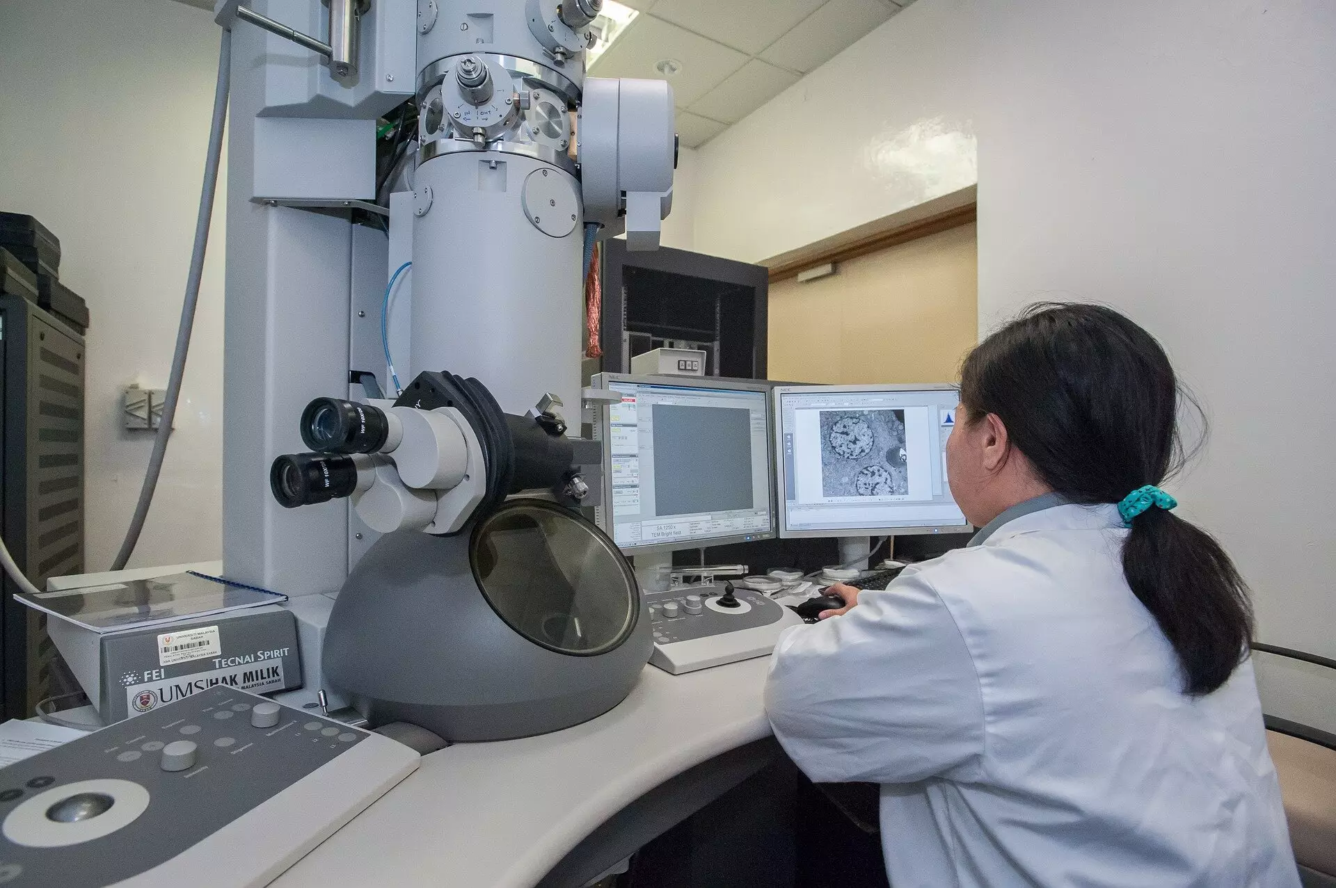
In an exciting leap for scientific research, a collaborative team of researchers from Trinity College Dublin and IDES Ltd. has unveiled a groundbreaking imaging technique that stands to transform various fields, from medicine to materials science. By leveraging advanced microscopy technology, they have introduced a method that significantly reduces both imaging time and radiation exposure. This innovative approach holds promise, particularly for sensitive materials such as biological tissues, which are often negatively affected by traditional imaging methods.
The conventional method of using scanning transmission electron microscopes (STEMs) has been the backbone of electron microscopy for years. Typically, these devices work by directing a finely focused electron beam across a sample, painstakingly assembling images pixel by pixel. The process involves a fixed “dwell time” at each pixel, akin to using traditional film cameras that require equal exposure time for each section of an image. This uniform exposure can lead to excessive radiation, potentially damaging delicate biological structures and skewing results.
Event-Based Detection: A Game Changer
What sets this new imaging approach apart is its reliance on event-based detection rather than the traditional fixed exposure methodology. The innovative technique recognizes that the first electron detected at any given point offers substantial information necessary for imaging, while subsequent electron impacts yield diminished returns. The team adeptly harnessed this insight to devise a method that requires fewer electrons for equivalent—or even superior—image quality.
Essentially, this new technique allows researchers to “shut off” the illumination of the electron beam when sufficient imaging efficiency has been achieved. By minimizing the number of electrons employed, they not only preserve sample integrity but also enhance the overall imaging process. This pivot in how imaging is conducted may seem subtle, yet it marks a paradigm shift in microscopy.
The Technical Marvel Behind the Method
At the heart of this innovative imaging strategy lies a patented technology dubbed Tempo STEM, an amalgamation of two cutting-edge technologies. This tool integrates a sophisticated “beam blanker” that can temporarily inhibit the electron beam as soon as the necessary precision has been met for any measurement point within the sample. This nanosecond-level control is unprecedented and represents a significant advancement in the capabilities of electron microscopy.
Dr. Lewys Jones, a key figure in this research and an esteemed assistant professor at Trinity, asserts that such technological synergy allows scientists to approach microscopy with a fresh perspective. The capability to manipulate the electron beam in response to real-time events not only enhances image clarity but also conserves sample integrity, thereby aiding in yielding more accurate results.
Implications for Scientific Research
The ramifications of this research are profound. In an era where precision is key, reducing radiation exposure has become a pressing necessity, especially when dealing with delicate biological samples. Current methodologies often result in damage, rendering acquired images unreliable or misleading. With the introduction of this new imaging technique, researchers will be able to investigate biological structures with much less risk of causing harm, opening up new avenues for discovery in fields such as cellular biology and medical diagnostics.
As Dr. Jon Peters, the first author of the research, points out, many scientists may underestimate the potential damage caused by high-speed electrons, yet the risks are significant when investigating fragile biological specimens. This modified approach not only addresses the challenges of traditional methods but also encourages a re-evaluation of existing imaging practices across various research domains.
A Future of Enhanced Exploration
As the scientific community stands on the brink of change, this innovative approach to imaging via reduced radiation and time represents a beacon of potential. While conventional microscopy has served researchers well, it often leads to compromised results. By shifting the focus to a more efficient model, one that minimizes risk and maximizes information, this new imaging method could catalyze a new era of discovery, enabling breakthroughs in our understanding of both living systems and material structures.
With the momentum of this revolutionary finding gaining traction, the anticipation of further developments in this field is palpable. Such advancements not only enhance scientific methodologies but also promise to spur collaborations that might redefine how we interact with the microscopic world. The future looks bright as researchers embrace these innovative tools, collectively propelling science toward new frontiers of knowledge and exploration.

