Recent advancements in medical imaging have led to the development of an innovative technique that allows researchers to visualize organs and biological structures by rendering overlying tissues transparent. This significant breakthrough, achieved by a team at Stanford University, utilizes a food-safe dye in a reversible application, opening avenues for enhanced medical diagnostics. This article delves into the scientific principles behind this technique, its applications, and its implications for future medical practices.
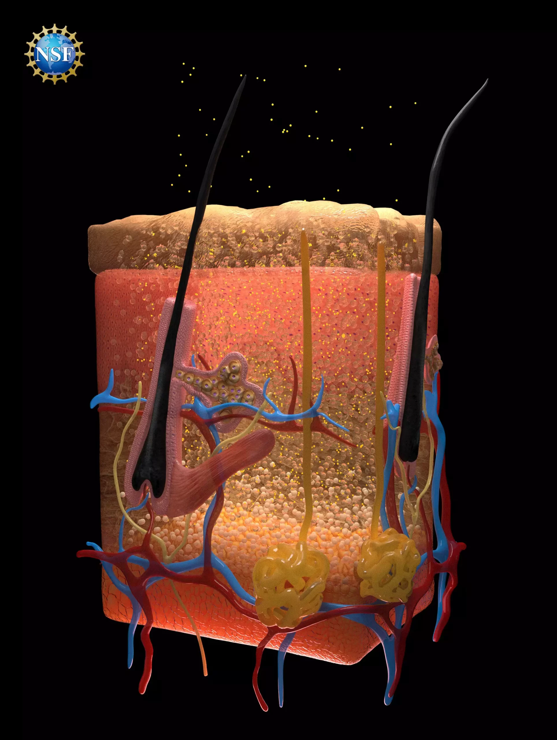
At the core of this innovative technology lies the manipulation of light interaction with biological tissues. Researchers focused on the concept of light scattering, which is a major factor preventing us from seeing through human bodies. Biological tissues—composed of fats, fluids, proteins, and cells—possess varying refractive indices, leading to a scattering effect that gives them an opaque appearance. The study titled “Achieving Optical Transparency in Live Animals with Absorbing Molecules,” published in the September 2024 issue of *Science*, details how researchers could potentially match these refractive indices with sophisticated dye solutions, enabling light to travel smoothly through tissues.
The team hypothesized that specific dyes could absorb light effectively while simultaneously facilitating its uniform passage through tissues. After extensive exploration, they identified a well-known food dye—tartrazine, commonly recognized as FD & C Yellow 5—whose molecular structure was ideal for this purpose. Tartrazine’s ability to align the refractive indices of different biological materials was the key to transforming solid organs into transparent structures, thus allowing for unprecedented levels of observation.
The researchers began their practical experiments with non-living tissue, utilizing thin slices of chicken breast. They observed that, as they increased the concentration of tartrazine, the refractive index of the surrounding fluid adjusted until it matched that of the muscle proteins. This manipulation resulted in the transparency of the tissue, reinforcing their initial hypothesis. Building on this successful experiment, the researchers applied the dye to living animal tissues, starting with gentle applications on the scalps of mice. This resulted in the visualization of blood vessels in the brain, and later, movement within the intestines, demonstrating the technique’s efficacy at microscopic scales.
Crucially, this procedure proved to be non-invasive and reversible. After the implementation of the dye, the researchers noticed that tissues reverted to their original opacity shortly after rinsing, and any excess dye was expelled naturally by the body within 48 hours. Such findings suggest a promising safety profile for potential therapeutic applications.
The implications of this optical transparency method could be profound. Beyond simply rendering tissues transparent for imaging, it opens doors for practical medical applications. For instance, this technology could assist in visualizing blood vessels more clearly for procedures such as blood draw or administering medications. Furthermore, it could enhance laser-based therapies by allowing penetrating light for better treatment of cancerous and precancerous tissues traditionally restricted to superficial areas.
Dr. Guosong Hong, the Stanford University assistant professor who played a pivotal role in this research, indicated that this technique could revolutionize early detection and treatment protocols across various medical fields. By improving light penetration, these therapies may yield more effective outcomes in treating conditions that necessitate precise targeting of affected tissues.
This groundbreaking work may set the stage for an entirely new field blending dye chemistry and optical physics with medicine. Researchers aim to refine and expand the applicability of their findings, exploring different dyes and their interactions with biological tissues. Historically, tools like the ellipsometer—commonly associated with semiconductor manufacturing—demonstrate the importance of interdisciplinary approaches in scientific research.
As optics continues to evolve, collaborating with biologists and medical professionals will be crucial in defining specific applications and achieving clinical integration of these techniques. This research not only emphasizes the importance of innovative thinking but also demonstrates the profound impact of combining knowledge from various scientific realms.
The quest for clearer images of organ systems and biological structures has taken an exhilarating turn with the advent of this new transparency technique. The research conducted at Stanford University highlights the potential of food-safe dyes in rendering biological tissues transparent. This advancement holds promising implications for the fields of diagnostics and treatment, which may one day enable faster, safer, and more accurate medical interventions. As this research progresses, continued exploration of the intersection between optics and biology will undoubtedly lead to further dramatic enhancements in medical imaging technologies.